
Chronic whiplash-associated disorders (WAD) can cause long-term neck pain, headaches, and cognitive issues, often following motor vehicle accidents. Traditional imaging like X-rays often fails to detect abnormalities, leading researchers to focus on biomarkers – measurable biological indicators of injury severity and treatment response.
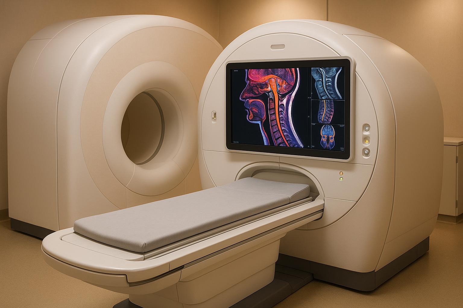
MRI (Magnetic Resonance Imaging) is emerging as an effective tool for identifying these biomarkers, offering detailed insights into soft tissue damage, muscle composition, inflammation, and nerve function. Advanced MRI techniques, such as Diffusion-weighted imaging (DWI) and functional MRI (fMRI), help detect subtle structural and functional changes in tissues that are often missed by conventional imaging.
Recent studies highlight muscle fat infiltration (MFI) as a promising biomarker for chronic WAD, linking it to higher pain levels and delayed recovery. These findings are helping clinicians develop more precise, personalized treatment plans, track progress, and improve patient care. However, challenges like high costs, patient accessibility, and insurance limitations remain. Future research aims to refine biomarkers further and explore advanced imaging technologies like upright MRI for better diagnostic accuracy.
MRI Methods for Chronic Whiplash Research
Main MRI Types for Chronic WAD Studies
Researchers rely on several advanced MRI techniques to identify biomarkers in patients with chronic whiplash.
Diffusion-weighted imaging (DWI) tracks the movement of water molecules in tissues. Under normal conditions, water moves freely, but damaged or inflamed tissues restrict this movement. In chronic whiplash cases, DWI can detect subtle changes in the spinal cord, brainstem, and surrounding tissues – changes that might not show up on standard MRI scans. This allows for the identification of microscopic structural damage that can persist long after the initial injury.
Morphological MRI focuses on changes in muscle composition, ligament thickness, and nerve root compression, all of which can correlate with a patient’s symptoms. For chronic whiplash-associated disorders, this technique can reveal muscle atrophy, scar tissue, and even shifts in the natural curvature of the cervical spine.
Phase-contrast imaging for cerebrospinal fluid (CSF) flow evaluates how CSF moves around the brain and spinal cord. Whiplash injuries can disrupt these flow patterns, potentially leading to headaches, dizziness, and cognitive issues. By measuring CSF flow velocity and volume, researchers can identify abnormalities that may explain why some patients experience chronic symptoms while others recover more quickly.
Functional MRI (fMRI) maps changes in brain activity associated with chronic pain. This technique provides valuable insights into central sensitization and how pain signals are amplified in the brain.
These specialized MRI methods provide data that not only advance research but also directly influence patient care.
How MRI Measurements Work in U.S. Clinical Settings
In chronic whiplash research, MRI measurements rely on precise quantification techniques that translate seamlessly into clinical practice. Even small changes – measured in millimeters – can hold significant clinical value, especially when evaluating muscle atrophy or ligament thickening.
Signal intensity measurements are reported in standardized units, allowing clinicians to compare results across patients and over time. Higher intensity often points to inflammation or swelling, while lower intensity may indicate fatty infiltration or fibrosis. Diffusion measurements are quantified using the apparent diffusion coefficient (ADC), which is measured in square millimeters per second. Deviations from normal ADC values can signal tissue injury.
CSF flow measurements are expressed in milliliters per minute. In clinical settings, abnormal flow rates often correlate with symptom severity, providing measurable data to guide treatment decisions and monitor progress.
These standardized measurements are easily integrated into electronic health records across U.S. healthcare systems, ensuring consistent communication among specialists and supporting coordinated care.
Using MRI in U.S. Chiropractic Clinics
Advanced MRI techniques are increasingly shaping treatment strategies in U.S. chiropractic clinics. Many clinics work closely with imaging centers and radiologists to obtain detailed MRI scans that inform their treatment plans.
Patient-focused imaging protocols are now the norm. Clinics often schedule MRI appointments with consideration for patients’ pain levels and mobility challenges. This might involve shorter scan sequences, the use of supportive positioning aids, and flexible scheduling to minimize discomfort during the imaging process.
For example, Portland Chiropractic Group emphasizes a team-based approach to interpreting MRI results. Instead of relying solely on radiologists’ reports, the clinical team reviews imaging findings alongside patient symptoms and functional assessments. This collaborative effort helps identify the biomarkers most relevant to each patient’s unique complaints, enabling more precise and effective treatment plans.
Insurance coverage for MRI studies related to chronic whiplash is widely available when clinical criteria are met. Detailed documentation of biomarker findings is critical for demonstrating the medical necessity of these studies, ensuring that patients can access advanced imaging when needed.
Integrating MRI findings into care has been particularly impactful. Chiropractors can adjust their techniques based on structural biomarkers, while partnering physicians may prescribe targeted therapies to address inflammation or nerve dysfunction revealed by imaging. This multidisciplinary approach allows for highly personalized treatment plans for chronic whiplash patients.
Recent MRI Study Results on Chronic Whiplash
What Recent Studies Show
Recent research using advanced MRI techniques sheds light on chronic whiplash-associated disorders (WAD), offering both new insights and ongoing challenges. One key discovery is the role of muscle fat infiltration (MFI) as a potential biomarker. A study involving 79 individuals with chronic WAD found significantly higher levels of fat infiltration in key cervical extensor muscles compared to healthy individuals[1][2].
Long-term studies also reveal that MFI in neck muscles increases between 3 and 6 months after injury. This increase is closely linked to higher levels of pain and posttraumatic stress during recovery[1][2].
In an effort to broaden diagnostic tools, Uhrenholt et al. conducted a feasibility study using advanced MRI techniques like quantitative diffusion-weighted imaging and cerebrospinal fluid flow measurements. However, they found no significant differences between chronic whiplash patients and healthy controls[1][2]. While these methods may need further refinement, the findings highlight MFI as a promising focus for diagnosing and understanding chronic WAD. This progress equips clinicians with better tools to fine-tune their diagnostic and treatment approaches.
What This Means for U.S. Doctors and Patients
For U.S. clinicians, the validation of MFI as a reliable biomarker offers a more objective way to diagnose chronic WAD. By assessing MFI, healthcare providers – such as chiropractors and physical therapists – can identify patients at higher risk for prolonged symptoms and tailor treatment plans accordingly. This approach not only aids early intervention but also allows professionals to track muscle changes over time, improving long-term care for patients dealing with chronic whiplash.
Clinical Uses and Next Steps
How MRI Helps Create Custom Treatment Plans
MRI scans play a key role in shaping personalized treatment plans by measuring muscle fat infiltration (MFI) in cervical extensor muscles [1]. At Portland Chiropractic Group, practitioners use these findings to adjust the intensity of treatments and create targeted muscle rehabilitation exercises. The insights from MRI scans allow them to address the specific level of muscle deterioration each patient experiences.
When scans show significant MFI in cervical muscles, chiropractors can offer patients a clearer understanding of their condition’s severity and establish realistic recovery timelines. This data-driven approach not only helps tailor treatment intensity but also tracks progress over time. Follow-up MRIs are essential for confirming whether treatments are successfully slowing or reversing muscle deterioration.
This patient-specific strategy underscores the commitment to improving chronic whiplash care through advanced imaging techniques. As research progresses, new opportunities to enhance these clinical applications continue to emerge.
Where Research Should Go Next
The future of chronic whiplash diagnosis and treatment lies in pushing the boundaries of imaging technology and clinical studies. One promising area is advanced spinal cord imaging. By incorporating Magnetization Transfer sequences, as recommended by the Spinal Cord Toolbox, researchers may uncover white matter lesions that current imaging methods overlook [3].
Another area of interest is upright MRI technology, which could provide a more accurate picture of structural changes. Supine MRI scans often miss abnormalities, such as tonsillar ectopia, that are only visible when the patient is upright. Research using upright MRI could shed light on these hidden contributors to chronic whiplash symptoms.
Beyond the spine, brain imaging techniques hold potential for early intervention strategies. Functional MRI studies targeting hippocampal activity could revolutionize how and when treatments are delivered. Dr. Apkarian explains:
"Now that we know there is this critical time period when this happens, we can focus our treatment efforts at this early stage to prevent chronic pain rather than try to cure it." [4]
He also suggests that future treatments could involve modulating hippocampal activity through pharmacological means or neuromodulation [4].
Large-scale, practice-based studies are another essential step. While much of the current research happens in controlled lab environments with small patient groups, expanding these studies to include hundreds of patients across multiple clinics would provide the robust data needed to refine diagnostic criteria and treatment protocols [3].
Additionally, future research must address confounding factors that impact MRI interpretations and patient outcomes. Variables like pain medication, depression, and other health conditions can complicate findings. Carefully controlled studies will help clarify these influences, leading to more precise clinical guidance [5].
These research directions aim to improve diagnostic accuracy and enable more personalized treatments. Beyond whiplash, the findings could extend to managing other chronic pain conditions, such as those resulting from sports injuries, workplace accidents, or degenerative diseases. This could pave the way for broader advancements in pain management across various medical specialties.
sbb-itb-ed556b0
Benefits and Drawbacks of MRI Biomarker Testing in Chronic Whiplash
MRI Benefits vs. Limitations Comparison Table
MRI biomarker testing offers both opportunities and challenges when diagnosing and managing chronic whiplash. Here’s a side-by-side look at its benefits and limitations:
| Benefits | Limitations |
|---|---|
| Non-invasive imaging – Delivers detailed views of soft tissues without surgery or harmful radiation | High cost – MRI scans are pricey, and multiple sessions may be needed for thorough assessments |
| Objective measurement – Tracks muscle fat infiltration (MFI) in cervical extensor muscles, offering data beyond patient-reported symptoms | Limited validated biomarkers – No standardized thresholds yet for a definitive chronic whiplash diagnosis |
| Treatment customization – Enables tailored rehabilitation plans and muscle-specific exercises based on tissue changes | Time-intensive process – MRI protocols can take a long time, complicating scheduling in busy clinics |
| Progress tracking – Follow-up scans help monitor whether treatments are slowing or reversing muscle deterioration | Patient limitations – Issues like claustrophobia, metal implants, or difficulty staying still can prevent some patients from completing scans |
| Early intervention potential – Advanced imaging techniques, such as Magnetization Transfer, may detect early-stage changes | Insurance coverage gaps – Advanced MRI protocols may not be fully covered, often requiring pre-authorization |
| Comprehensive assessment – Modern MRI can simultaneously examine muscles, ligaments, the spinal cord, and brain structures | Interpretation complexity – Distinguishing injury-related changes from age-related variations demands specialized expertise |
Balancing these benefits and limitations is essential for making informed decisions in clinical practice. MRI biomarker testing can provide valuable insights, but practical challenges like cost, patient accessibility, and interpretation hurdles remain significant.
As research progresses, the development of validated biomarkers and standardized protocols may help address some of these limitations. This could make MRI biomarker testing a more routine tool in chronic whiplash care, offering a more personalized approach to diagnosis and treatment.
Conclusion: How MRI Can Improve Chronic Whiplash Care
MRI technology is changing the game when it comes to diagnosing and treating chronic whiplash-associated disorders. Studies have shown that muscle fat infiltration (MFI) in the cervical extensor muscles provides clear, measurable evidence to differentiate chronic whiplash patients from those without the condition. For instance, a study involving 79 individuals with chronic WAD revealed significantly higher levels of fat infiltration in key cervical muscles compared to healthy participants[6].
"There is substantial evidence supporting the use of muscle fat infiltration (MFI) as a biomarker to distinguish chronic whiplash patients from healthy controls." – Julia Evans and Michael Fishman[1]
These findings are driving a shift toward more precise and individualized care. Instead of relying solely on subjective patient reports, MRI biomarkers offer concrete data that can guide treatment decisions and monitor progress. Advances in MRI techniques are also opening up new diagnostic opportunities.
At Portland Chiropractic Group, this research reinforces our commitment to evidence-based care. Dr. Brett Weaver and our team use MRI biomarker data to tailor rehabilitation plans, whether it’s through targeted Graston technique, specialized dry needling, or customized functional training exercises. When patients see the MRI results, it not only validates their symptoms but also builds trust in our transparent and patient-focused approach. By addressing the specific needs of affected tissues, these personalized treatments aim to deliver better outcomes. As research continues to refine biomarkers and establish standardized protocols, MRI is poised to become an essential tool for developing more effective and targeted treatments for chronic whiplash patients.
FAQs
How do advanced MRI techniques like DWI and fMRI help diagnose and treat chronic whiplash-associated disorders more effectively than traditional imaging?
Advanced MRI methods like diffusion-weighted imaging (DWI) and functional MRI (fMRI) offer deeper insights into the effects of chronic whiplash compared to traditional imaging techniques. DWI excels at detecting subtle nerve and tissue damage by creating detailed three-dimensional images, while fMRI tracks brain activity linked to pain and injury, shedding light on how the nervous system is involved.
Unlike conventional MRI, which focuses primarily on bones and major soft tissue injuries, these advanced techniques reveal physiological changes that are often missed. This enhanced clarity allows for more precise diagnoses and supports personalized treatment plans aimed at managing chronic pain more effectively and improving recovery outcomes.
How does muscle fat infiltration (MFI) act as a biomarker for chronic whiplash, and how can it guide personalized treatment?
Muscle fat infiltration (MFI) describes the accumulation of fat within neck muscles, a phenomenon commonly seen in individuals dealing with chronic whiplash. This buildup is often associated with lingering symptoms and slower recovery, making it an important indicator for identifying patients who may need additional care.
MRI scans allow doctors to assess MFI levels, offering a clearer picture of the muscle changes involved. With this insight, healthcare providers can design tailored treatment plans focused on muscle rehabilitation and reducing fatty deposits. This approach not only supports better recovery but also helps in managing chronic pain more effectively.
What challenges do doctors face when using MRI to identify biomarkers for chronic whiplash, and how could these be resolved in the future?
One of the biggest hurdles in using MRI to address chronic whiplash lies in its difficulty detecting subtle physiological changes often associated with the condition. Adding to the challenge is the absence of standardized biomarkers, which complicates consistent diagnosis and tracking of a patient’s progress. Furthermore, clinical validation is still underway, requiring additional research to ensure dependable results.
Looking ahead, advancements in imaging technology, the creation of standardized protocols, and larger-scale research studies hold the potential to greatly enhance MRI’s accuracy and effectiveness in diagnosing and managing chronic whiplash. These developments could pave the way for improved treatment approaches and better outcomes for individuals suffering from chronic pain due to whiplash injuries.
Related Blog Posts
- The Science Behind Why Chiropractic Care is the Top Treatment of Choice for Whiplash Associated Disorders
- Why Is Fatty Infiltration in the Multifidi Muscle Important for a Chronic Low Back Pain Prognosis?
- Effectiveness of Articular and Neural Mobilization for Managing Cervical Radicular Pain: A Systematic Review With Network Meta-Analysis
- Association between cervical MRI findings and patient-reported severity of headache in patients with persistent neck pain: a cross-sectional study

Comments are closed